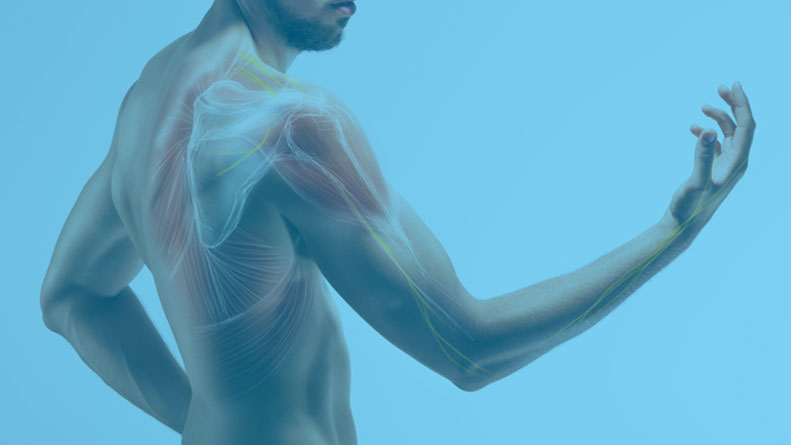
Understanding A Shoulder Injury
Shoulder Anatomy
The shoulder is the most flexible joint in the body, enabling a wide range of movements including forward flexion, abduction, adduction, external rotation, internal rotation, and 360-degree circumduction.
Thus, the shoulder joint is considered the most insecure joint of the body but the support of ligaments, muscles, and tendons function to provide the required stability.
Bones
Soft Tissues
Ligaments
Muscles & Tendons
Nerves & Blood Vessels
Bones
Bones of the Shoulder
The shoulder is a ball and socket joint made up of three bones, namely the humerus, scapula, and clavicle.
The end of the humerus or upper arm bone forms the ball of the shoulder joint. An irregular shallow cavity in the scapula called the glenoid cavity forms the socket for the head of the humerus to fit in. The two bones together form the glen humeral joint, which is the main joint of the shoulder.
The scapula is a flat triangular-shaped bone that forms the shoulder blade. It serves as the site of attachment for most of the muscles that provide movement and stability to the joint. The scapula has four bony processes – acromion, spine, coracoid, and glenoid cavity. The acromion and coracoid process serve as places for attachment of the ligaments and tendons.
The clavicle bone or collarbone is an S-shaped bone that connects the scapula to the sternum or breastbone. It forms two joints: the acromioclavicular joint, where it articulates with the acromion process of the scapula, and the sternoclavicular joint where it articulates with the sternum or breast bone. The clavicle also forms a protective covering for important nerves and blood vessels that pass under it from the spine to the arms.
Soft Tissues
Soft Tissues of the Shoulder
The ends of all articulating bones are covered by smooth tissue called articular cartilage which allows the bones to slide over each other without friction, enabling smooth movement. Articular cartilage reduces pressure and acts as a shock absorber during movement of the shoulder bones.
Extra stability to the glenohumeral joint is provided by the glenoid labrum, a ring of fibrous cartilage that surrounds the glenoid cavity. The glenoid labrum increases the depth and surface area of the glenoid cavity to provide a more secure fit for the half-spherical head of the humerus.
Ligaments
Ligaments of the Shoulder
Ligaments are the thick strands of fibers that connect one bone to another. The ligaments of the shoulder joint include:
- Coraco-clavicular ligaments: These ligaments connect the collarbone to the shoulder blade at the coracoid process.
- Acromio-clavicular ligament: This connects the collarbone to the shoulder blade at the acromion process.
- Coraco-acromial ligament: It connects the acromion process to the coracoid process.
- Glenohumeral ligaments: A group of 3 ligaments that form a capsule around the shoulder joint, and connect the head of the arm bone to the glenoid cavity of the shoulder blade. The capsule forms a water-tight sac around the joint. Glenohumeral ligaments play a very important role in providing stability to the otherwise unstable shoulder joint by preventing dislocation.
Muscles & Tendons
Muscles of the Shoulder
The rotator cuff is the main group of muscles in the shoulder joint and is comprised of four muscles. The rotator cuff forms a sleeve around the humeral head and glenoid cavity, providing additional stability to the shoulder joint while enabling a wide range of mobility.
The deltoid muscle forms the outer layer of the rotator cuff and is the largest and strongest muscle of the shoulder joint.
Tendons of the Shoulder
Tendons are strong tissues that join muscle to bone allowing the muscle to control the movement of the bone or joint. Two important groups of tendons in the shoulder joint are the biceps tendons and rotator cuff tendons.
Bicep tendons are the two tendons that join the bicep muscle of the upper arm to the shoulder. They are referred to as the long head and short head of the bicep.
Rotator cuff tendons are a group of four tendons that join the head of the humerus to the deeper muscles of the rotator cuff. These tendons provide more stability and mobility to the shoulder joint.
Nerves & Blood Vessels
Nerves of the Shoulder
Nerves carry messages from the brain to muscles to direct movement (motor nerves) and send information about different sensations such as touch, temperature, and pain from the muscles back to the brain (sensory nerves). The nerves of the arm pass through the shoulder joint from the neck.
These nerves form a bundle at the region of the shoulder called the brachial plexus. The main nerves of the brachial plexus are the musculocutaneous, axillary, radial, ulnar, and median nerves.
Blood Vessels of the Shoulder
Blood vessels travel along with the nerves to supply blood to the arms. Oxygenated blood is supplied to the shoulder region by the subclavian artery that runs below the collarbone. As it enters the region of the armpit, it is called the axillary artery and further down the arm, it is called the brachial artery. The main veins carrying de-oxygenated blood back to the heart for purification include:
- Axillary vein: this vein drains into the subclavian vein
- Cephalic vein: this vein is found in the upper arm and branches at the elbow into the forearm region. It drains into the axillary vein.
- Basilic vein: this vein runs opposite the cephalic vein, near the triceps muscle. It drains into the axillary vein.
Conditions
Shoulder Pain
Acromioclavicular Injuries
Clavicle Fracture
Dislocation/Subluxation
Frozen Shoulder
Labral Tear/SLAP Tear
Rotator Cuff Tear
Shoulder Impingement
Shoulder Pain
What is Shoulder Pain?
Pain in the shoulder suggests a shoulder injury which is more common in athletes participating in sports such as swimming, tennis, pitching, and weightlifting. The injuries are caused due to the over usage or repetitive motion of the arms.
In addition to pain, shoulder injuries also cause stiffness, restricted movements, difficulty in performing routine activities, and a popping sensation.
Causes of Shoulder Pain
Some of the common shoulder injuries that cause pain and restrict the movement of shoulders include sprains and strains, dislocations, tendinitis, bursitis, rotator cuff injury, fractures, and arthritis.
- Sprains and strains: A sprain is the stretching or tearing of ligaments (tissues that connect adjacent bones in a joint). It is a common injury and usually occurs when you fall or suddenly twist. A strain is stretching or tearing of muscle or tendon (tissues that connect muscle to bone). It is common in people participating in sports. Strains are usually caused by the twisting or pulling of the tendons.
- Dislocations: A shoulder dislocation is an injury that occurs when the end of the bone is forced out of its position. It is often caused by a fall or direct blow to the joint while playing a contact sport.
- Tendinitis: It is an inflammation of a tendon, which is fibrous collagen tissue that connects muscles to bone. It occurs because of injury or overuse.
- Bursitis: It is an inflammation of a fluid-filled sac called bursa that protects and cushions your joints. Bursitis can be caused by chronic overuse, injury, arthritis, gout, or infection.
- Rotator cuff injury: The rotator cuff consists of tendons and muscles that connect your shoulder blade to your humerus (arm bone). Rotator cuff muscles allow you to move your arm up and down. Rotator cuff injuries often cause a decreased range of motion.
- Fractures: A fracture is a break in the bone that commonly occurs because of injury, such as a fall or a direct blow to the shoulder.
- Arthritis: Osteoarthritis is the most common type of shoulder arthritis, characterized by progressive wearing away of the cartilage of the joint.
Diagnosis of Shoulder Pain
Shoulder conditions should be evaluated by Dr. Gregory DiFelice for a proper diagnosis and treatment. A detailed medical history and physical examination of the knee are crucial for the diagnosis. Dr. Gregory DiFelice may also conduct diagnostic imaging studies such as X-rays, MRI scans, CT scans, and ultrasound, to help visualize the bony structures and soft tissues of the shoulder.
Treatment for Shoulder Pain
Treatment options depend upon the underlying cause responsible for shoulder pain. Early treatment is necessary to prevent serious shoulder injuries. The immediate mode of treatment recommended for shoulder injuries is rest, ice, compression, and elevation (RICE). Dr. Gregory Difelice may recommend the use of over-the counter medications, cortisone injections, and physical therapy based off your injuries.
For more information, please contact our office to schedule your appointment today.
Acromioclavicular Injuries
What is an Acromioclavicular (AC) Joint Injuries?
The acromioclavicular (AC) joint is the connection between the end of your clavicle (collar bone) and the roof of your shoulder (acromion). This joint complex is comprised of the acromioclavicular ligament, and the coracoclavicular ligament. While this joint complex is strong, its location makes it susceptible to direct trauma injuries. Overloading of this joint can result in disruption of ligament fibers, causing the end of the clavicle to shift, resulting in pain. This is also called a “separated” shoulder. Dr. Gregory DiFelice specializes in diagnosing and treating AC joint injuries.
Causes of AC Joint Injuries
An AC joint separation is often the result of direct trauma to the shoulder itself, frequently from a fall. These types of injuries are frequently seen in males in their 20s, often from contact sports, such as ice hockey, rugby, and football. Less frequently, these injuries can be the result of a call onto an outstretch arm or elbow. Injuries to the AC joint can be graded as types 1 through 6, depending on the severity of the injury.
Type 1: Mild injury, spraining of the acromioclavicular (AC) ligament, however, AC joint is intact
Type 2: Low-grade injury, tearing of the AC ligament, however, the corcacoclavicular (CC) ligament is intact. There is mild shifting of the clavicle up but not dislocated.
Type 3: Moderate injury, with complete tearing of the AC and CC ligaments with complete dislocation of the AC joint
Type 4, 5, and 6: Severe injuries, with complete tearing of the AC and CC ligaments with displacement of the clavicle, either backwards, forwards, or upwards.
Symptoms of AC Joint Injuries
Patients with an AC joint separation will have symptoms of shoulder pain after a fall, with decreased range of motion. Patients may experience bruising, swelling, or a bump on the top of their shoulder. Weakness and difficulty with raising your arm forwards or to the side is common.
Diagnosis of AC Joint Injuries
Dr. Gregory DiFelice will make a diagnosis of an AC joint separation based on your symptoms, medical history, and by performing a physical examination of your shoulder. Other diagnostic tests such as X-rays and MRI scans may be ordered. X-rays are useful to evaluate the degree of separation of the AC joint. An MRI scan is done to help view the images of soft tissues better and to ensure that no other structures are involved.
Treatment of AC Joint injuries
Treatment options may include non-surgical and surgical treatment. Non-surgical treatment is frequently recommended for type 1 and 2 injuries. This may consist of rest, ice, and physical therapy. Based off of your pain, Dr. Gregory DiFelice may sometimes recommend the use of sling. Physical therapy will include several weeks of range of motion and strengthening exercises.
Generally, surgery is considered in patients with type 3-6 AC joint injuries. Using his Preservation First™ approach, Dr. Gregory DiFelice will assess if you will need an AC joint repair or AC joint reconstruction. This will be based off of your age, injury, tissue quality, and injury pattern. Repairing an AC joint separation will involve passing a button placed onto a thick, sturdy stitch through a small drill hole in your clavicle and your coracoid process. A second button will then be used to tie down this construct and bring the two ends of the AC joint closer together. If the injury pattern is too extensive Dr. Gregory DiFelice may need to reconstruct the damaged ligament using a tissue graft. Following an AC joint separation repair or reconstruction, you will be placed into sling to limit your range of motion. A rehabilitation program will be started that helps you resume a wider range of activities. Usually, a complete recovery may take about 6 months depending on your injury pattern.
For more information, please contact our office to schedule your appointment today.
Clavicle Fracture
What is a Clavicle Fracture?
The clavicle, also known as the collarbone, is an “S” shaped bone that serves as the only bony connection between your arm and your trunk. Due to its location, the clavicle is one of the most frequently fractured bones. Clavicle fractures are a very common sports injury seen in people who are involved in contact sports such as football and martial arts as well as impact sports such as motor racing. Dr. Gregory DiFelice specializes in diagnosing and treating clavicle fractures.
Causes of Clavicle Fracture
Fractures of the clavicle are from direct trauma to the shoulder. Traffic accidents and sports injuries account for most of these injuries. Falls onto an outstretch hand can cause a clavicle fracture, but these are less frequent. Fractures of the clavicle are classified based on the location of the injury.
Group 1: Fractures that occur in the middle of the bone, also known as midshaft fracture.
Group 2: Fractures that occur closer to the shoulder, also known as lateral or distal fracture.
Group 3: Fractures that occur closer to the neck, also known as medial or proximal fracture.
Symptoms of Clavicle Fractures
Patients with a clavicle fracture will have symptoms of shoulder and collarbone pain after a fall, with decreased range of motion. Patients may experience bruising, swelling, or a bump over their collarbone. Weakness and difficulty with raising your arm forwards or to the side is common.
Diagnosis of Clavicle Fractures
Dr. Gregory DiFelice will make a diagnosis of a clavicle fracture based on your symptoms, medical history, and by performing a physical examination of your shoulder. Other diagnostic tests such as X-rays, CT scans, and MRI scans may be ordered. X-rays are useful to evaluate the degree of displacement of the fractured fragments. A CT scan may be ordered to help evaluate the fracture pattern and help with surgical planning. An MRI scan is done to help view the images of soft tissues better and to ensure that no other structures are involved.
Treatment of Clavicle Fractures
Treatment options may include non-surgical and surgical treatment. Non-surgical treatment is recommended if the fracture bone has not shifted, or is “nondisplaced.” This may consist of rest, ice, and the use of a sling. Once the bone has healed, Dr. Gregory DiFelice may recommend PT to help with range of motion.
Generally, surgery is considered in patients displaced fractures. The primary surgical option for clavicle fractures is an open reduction and internal fixation, or ORIF. To perform this, Dr. Gregory DiFelice will realign the bone ends and hold them in place with the use of hardware, including plates and screws. Following an ORIF of the clavicle, you will be placed into sling to limit your range of motion. A rehabilitation program will be started that helps you resume a wider range of activities. Generally, the surgical hardware will remain in place, however, if they cause any irritation, they can be removed after the fracture heals. Usually, a complete recovery may take about 3 to 6 months depending on your injury pattern.
For more information, please contact our office to schedule your appointment today.
Dislocation/Subluxation
What is Shoulder Dislocation/Subluxation?
The shoulder is a ball-and-socket joint made up of three bones, namely the humerus (arm bone), scapula (shoulder blade), and clavicle (collarbone). The “ball” portion of the joint is the end of the humerus bone. This moves along the shallow dish, or socket, called the glenoid which is part of the scapula. Together these two bones make up the glenohumeral joint. Direct contact to this joint can force the humerus out of its normal position. Gregory DiFelice specializes in diagnosing and treating shoulder dislocations/subluxations.
Symptoms of Shoulder Dislocation
Most common symptoms of shoulder dislocation are pain and shoulder joint instability. Other symptoms such as swelling, numbness, and bruising may occur. At times, it may cause a tear in the ligaments or tendons of the shoulder as well as nerve damage.
Causes of a Shoulder Dislocation/Subluxation
Playing overhead and contact sports, or falling out onto an outstretch arm can cause the humerus to slide out of the joint. The dislocation might be a partial dislocation, or a subluxation or a complete dislocation causing pain and shoulder joint instability. The shoulder joint often dislocates in the forward direction, anteriorly, however, it may also dislocate in the backward, or posteriorly.
Symptoms of Shoulder Dislocation
Most common symptoms of shoulder dislocation are pain and shoulder joint instability. Other symptoms such as swelling, numbness, and bruising may occur. At times, it may cause a tear in the ligaments, labrum, or tendons of the shoulder as well as nerve damage. You may notice a visible dip on the top of your shoulder and see a bump in the front of your shoulder.
Diagnosis of Shoulder Dislocation
Dr. Gregory DiFelice will make a diagnosis of a shoulder dislocation/subluxation based on your symptoms, medical history, and by performing a physical examination of your shoulder. Other diagnostic tests such as X-rays, CT scans, and MRI scans may be ordered. X-rays are useful to evaluate if the dislocation is anterior or posterior. An MRI scan is done to help view the images of soft tissues better and to evaluate if there was tearing to labrum.
Treatments for Shoulder Dislocation
The condition is treated by a process called closed reduction which involves placing the ball of the upper arm back into the socket. Following this, the shoulder will be immobilized using a sling for several weeks. Ice may be applied over the area for 3-4 times a day. An MRI will be ordered to evaluate the soft tissue of the shoulder and rule out tearing to the labrum. Ice may be applied over the area for 3-4 times a day. Rehabilitation exercises may be started to restore range of motion, once the pain and swelling decrease.
For more information, please contact our office to schedule your appointment today.
Frozen Shoulder
What is Frozen Shoulder?
Frozen shoulder, also called adhesive capsulitis, is a condition characterized by pain and loss of motion in the shoulder joint. This condition rarely affects both shoulders. It is more common in older adults aged between 40 and 60 years and is more common in women than men. Frozen shoulder is often associated with other medical conditions, however, can also come from direct injuries. Frozen shoulder is usually self-limiting, however, in some cases requires surgical treatments. Gregory DiFelice specializes in diagnosing and treating frozen shoulder.
Causes of Frozen Shoulder
Frozen shoulder is caused by inflammation of the ligaments holding the shoulder bones to each other. The shoulder capsule becomes thick, tight, and the stiff bands of tissue called adhesions, or scar tissue, may develop. Risk factors for developing frozen shoulder include shoulder injuries, shoulder surgeries, a shoulder immobilized in a sling for longer a period, and other disease conditions such as diabetes, hypothyroidism, hyperthyroidism, Parkinson’s disease, or cardiac disease.
Symptoms of Frozen Shoulder
Frozen shoulder may cause pain and stiffness and limit the movements of the shoulder. Frozen shoulder is commonly classified into the following three stages, based off your symptoms.
- Freezing Stage: The shoulder becomes increasingly painful and difficulty to raise above your head. This process generally takes about six to nine weeks.
- Frozen Stage: The shoulder continues to remain stiff, however, the pain is usually lessened. If untreated, this stage can last about four to six months and can make your daily activities quite difficult.
- Thawing Stage: The shoulder begins to “loosen” up allowing you to increase your range of motion. During this phase, your daily activities become easier. This stage generally takes six to twelve months.
Diagnosis of Frozen Shoulder
Dr. Gregory DiFelice will make a diagnosis of frozen shoulder based on your symptoms, medical history, and by performing a physical examination of your shoulder. Dr. Gregory DiFelice may also conduct diagnostic imaging studies such as X-rays, MRI scans, CT scans, and ultrasound, to help visualize the bony structures and soft tissues of the shoulder.
Treatment Options for Frozen Shoulder
Treatment options may include non-surgical and surgical treatment. Non-surgical treatment is usually the initial course of treatment for frozen shoulder. This option consists of rest, ice, non-steroidal anti-inflammatory medications, cortisone injections, and physical therapy. This may take several months of physical therapy and repeat cortisone injections to fully resolve. Most cases of adhesive capsulitis are self-limiting, however, and will resolve with time. If your symptoms persist after several months of conservative treatment, surgical options may need to be consider.
Generally, surgery is considered as a last resort for patients with persistent symptoms despite non-surgical treatment. Surgery is performed arthroscopically with a few small incisions. A capsular release will be performed by debriding and cauterizing existing scar tissue in the shoulder will then be debrided and cauterized. A rehabilitation program focused on aggressive range of motion will be started that helps you resume a wider range of activities. Usually, a complete recovery may take about 12 to 24 months.
For more information, please contact our office to schedule your appointment today.
Labral Tear/SLAP Tear
What is a Shoulder Labrum Tear/SLAP Tear?
The shoulder joint is a “ball and socket” joint that enables smooth gliding and thereby the movements of arms. However, it is inherently unstable because of the shallow socket of the glenoid. A soft rim of cartilage, called the labrum, lines the socket and deepens it so that it accommodates the head of the upper arm bone better. Overhead injuries to shoulder can result in tearing to this labrum, resulting in continued pain and instability. Dr. Gregory DiFelice specializes in diagnosing and treating labral tears.
Causes of Shoulder Labral Tear
Traumatic injury to the shoulder or overuse of shoulder, such as throwing or weightlifting, may cause a labral tear. The most common causes include falling onto an outstretched arm, reoccurring dislocations, or overhead activities. Overhead and contact sports may put you at a greater risk of developing labral tears. Labral tears are classified by their location. Common types of labral tears include anterior, posterior, inferior, and superior. A SLAP tear is a labral tears that is located in the superior labrum, that runs front to back, or anterior to posterior.
Symptoms of Shoulder Labral Tear
Shoulder labral tear injury may cause symptoms such as pain, catching or locking sensation, decreased range of motion, and joint instability. Athletes, such as pitchers, may report pain while throwing. Patients may note instability in the shoulder, particularly when raise their arm above their head.
Diagnosis of Shoulder Labral Tear
Dr. Gregory DiFelice will make a diagnosis of a labral tear based on your symptoms, medical history, and by performing a physical examination of your shoulder. Dr. Gregory DiFelice may also conduct diagnostic imaging studies such as X-rays and MRI scans, to help visualize the bony structures and soft tissues of the shoulder.
Treatments for Shoulder Labral Tear
Treatment options may include non-surgical and surgical treatment. Non-surgical options are commonly reserved for patients with simple labral tears who are not experiencing instability. These options include activity modification, rest, ice, non-steroidal anti-inflammatory medications, cortisone injections, and physical therapy. If your symptoms surgical options may need to be consider.
Generally, surgery is considered in patients persistent symptoms, despite trying conservative options. Surgery options include a labral repair, also known as a stabilization, or a debridement. This decision will be based off of your symptoms, age, tissue quality, level of activity, and injury pattern. This surgery will be performed arthroscopically with a few small incisions. If the labral tear is simple, then the tissue will be debrided and smoothed out. However, if there is an extensive tear, with complete separation of the labrum from the glenoid, than a repair will be performed. The displaced labral tissue will be stitched back to the glenoid and anchored with small, non-metallic anchors. Depending on the extent of your tear, this may require several anchors. For a labral debridement, you will be placed in a sling for comfort for the first week. Following a labral repair, or stabilization, you will be placed into sling for about 4 weeks to limit your range of motion. A rehabilitation program will be started that helps you resume a wider range of activities. Usually, a complete recovery may take about 6 months depending on your injury pattern.
For more information, please contact our office to schedule your appointment today.
Rotator Cuff Tear
What is a Rotator Cuff Tear?
The rotator cuff is the group of four muscles that come together as tendons in the shoulder joint to provide support and enable a wider range of motion. The rotator cuff consists of the supraspinatus, the infraspinatus, the teres minor, and the subscapularis. These muscle connect the scapula, or shoulder blade, to the humerus, or arm bone. Major injury to these tendons may result in tear of these tendons and the condition is called as rotator cuff tear. It is one of the most common causes of shoulder pain in middle aged adults and older individuals. Dr. Gregory DiFelice specializes in diagnosing and treating rotator cuff tears.
What are the Causes of Rotator Cuff Tears?
Rotator cuff tears result from pressure on the rotator cuff from part of the scapula, or shoulder blade, as the arm is lifted. It may occur with repeated use of arm for overhead activities, while playing sports, or during motor accidents. Bone spurs located on the undersurface of your shoulder, or the acromion, can cause wear down to the rotator cuff overtime, causing tearing. Smoking may be a risk factor for rotator cuff tears. In older patients, a fall onto an outstretched arm can cause tearing of the rotator cuff tendons. Rotator cuff tears can be partial or complete tears.
What are the Symptoms of Rotator Cuff Tears?
Rotator cuff tears can cause mild to severe pain depending on the degree of the tear. Patients may experience pseudoparalysis, or the inability to raise their arm. There may be a crackling sensation with certain movement. There may also be stiffness, swelling, loss of movement, and tenderness in the front of the shoulder.
How is a Rotator Cuff Tear diagnosed?
Dr. Gregory DiFelice will make a diagnosis of a rotator cuff tear based on your symptoms, medical history, and by performing a physical examination of your shoulder. Dr. Gregory DiFelice may also conduct diagnostic imaging studies such as X-rays and MRI scans, to help visualize the bony structures and soft tissues of the shoulder.
What are Treatment Options for Rotator Cuff Tears?
Treatment options may include non-surgical and surgical treatment. Non-surgical options are commonly reserved for patients with partial rotator cuff tears who are experiencing persistent weakness. These options include activity modification, rest, ice, non-steroidal anti-inflammatory medications, cortisone injections, and physical therapy. If your symptoms persist surgical options may need to be consider.
Generally, surgery is considered in patients persistent symptoms, despite trying conservative options. Surgery options include a rotator cuff repair or a debridement. This decision will be based off of your symptoms, age, tissue quality, level of activity, and injury pattern. This surgery will be performed arthroscopically with a few small incisions. If the rotator cuff tear is a partial tear, then the tissue will be debrided and smoothed out. However, if there is an extensive tear, with complete separation of the tendon to the bone, than a repair will be performed. The displaced tendon will be stitched back to the humeral head and anchored with small, non-metallic anchors. Depending on the extent of your tear, this may require several anchors. For a rotator cuff debridement, you will be placed in a sling for comfort for the first week. Following a rotator cuff repair, you will be placed into sling for about 4 weeks to limit your range of motion. A rehabilitation program will be started that helps you resume a wider range of activities. Usually, a complete recovery may take about 6 to 9 months depending on your injury pattern.
For more information, please contact our office to schedule your appointment today.
Shoulder Impingement
What is Shoulder Impingement?
Shoulder impingement is the condition of inflammation of the tendons of the shoulder joint. It is one of the most common causes of pain in the adult shoulder. The shoulder is a ‘ball-and-socket’ joint. A ‘ball’ at the top of the upper arm bone, or humerus, fits neatly into a ‘socket,’ called the glenoid, which is part of the shoulder blade or scapula. Pain is caused when the rotator cuff is compressed by the roof of the shoulder, or acromion, when lifting. Shoulder impingement is also call “Rotator Cuff Syndrome” or “swimmer’s shoulder.” Dr. Gregory DiFelice specializes in diagnosing and treating rotator cuff tears.
Causes of Shoulder Impingement
Impingement results from pressure on the rotator cuff from the acromion, or roof of the shoulder, as the arm is lifted. It is more likely to occur in young and middle-aged people who engage in physical activities that require repeated overhead arm movements. The pain may be due to a “bursitis” or inflammation of the bursa overlying the rotator cuff or a “tendonitis” of the cuff itself. In some circumstances, a partial tear of the rotator cuff may cause impingement pain.
Symptoms of Shoulder Impingement
Individuals with shoulder impingement may experience severe pain at rest and during activities, weakness of the arm, and difficulty in raising the hand overhead. Patients frequently note pain at night, when they attempt to sleep on the affected side. Patients may hear grinding or clicking as they raise their arm overhead. Patients with rotator cuff syndrome more commonly note pain, not weakness, with overhead motions.
Diagnosis of Shoulder Impingement
Dr. Gregory DiFelice will make a diagnosis of a rotator cuff syndrome based on your symptoms, medical history, and by performing a physical examination of your shoulder. Dr. Gregory DiFelice may also conduct diagnostic imaging studies such as X-rays and MRI scans, to help visualize the bony structures and soft tissues of the shoulder.
Treatment Options for Shoulder Impingement
Treatment options may include non-surgical and surgical treatment. Non-surgical options include activity modification, rest, ice, non-steroidal anti-inflammatory medications, cortisone injections, and physical therapy. If your symptoms surgical options may need to be consider.
Generally, surgery is considered in patients persistent symptoms, despite trying conservative options. The primary surgical options includes a subacromial decompression and debridement. This surgery will be performed arthroscopically with a few small incisions. Degenerative tissue will be removed from your shoulder, as well as any bone spurs which may be causing the impingement. After surgery, you will be placed in a sling for comfort for the first week. A rehabilitation program will be started that helps you resume a wider range of activities. Usually, a complete recovery may take about 3 to 9 months depending on your injury pattern.
For more information, please contact our office to schedule your appointment today.